X-ray Absorption Spectroscopy
From Elucidating the Electronic Origin of Spectroscopic Signatures to Tracking Reaction Intermediates by In-Situ/Operando Approaches

X-ray absorption spectroscopy (XAS) is a highly sensitive and element-specific spectroscopy that provides detailed information on electronic structures, arising from different transitions, depending on the respective core electrons excited through the incident photon beam (K-, L-, M-edge XAS). Notably, XAS provides specific information at each edge, when focusing on the pre-edge, the near-edge structure (XANES or near-edge X-ray absorption fine structure (NEXAFS), when discussing soft X-rays) and the extended X-ray absorption fine structure (EXAFS), respectively.
Tracking Reactive Intermediates
Our group uses in situ or Operando XAS to detect key reaction intermediates. For instance, we (D. Lebedev et al.) obtained the spectroscopic signatures of the elusive Ru(V) and Ir(V)-Oxo intermediates, involved in the oxygen evolution reaction (EOR), a key process in water oxidation.[1,2] In some cases, the active sites are only a small fraction of the observed XAS signatures so that it is only possible to observe dormant species, even under Operando conditions. Therefore, developing state-of-the-art measurement schemes, i.e. gas switching and period averaging experiments, has enabled to track minute surface selective changes under reaction conditions. In that context, we (S. Docherty et al.) were able to detect surface redox dynamics in gold-zinc CO2 hydrogenation catalysts, [3] by amplification of subtle changes occurring at the surface, thus enabling to detect surface dynamic phenomena that correlates with the observed reactivity.

Identifying Surface Sites from “Augmented” XAS Libraries
Even though XAS-based characterization is crucial for understanding the nature of active sites of heterogenous catalysts, the majority of studies still rely on a limited number of solid-state references that may not provide relevant information about surface sites, which are likely associated with unusual coordination environments. We have therefore focused on establishing molecular libraries and developing machine learning approaches to capture the structure of surface sites. We (D. Trummer et al.) use Cr-based molecular libraries to relate observed spectroscopic signatures to specific electronic signatures based on computational approaches. This approach has enabled to carry out a quantitative analysis of the pre-edge region in Cr-K-edge XANES of the Phillips catalysts at various stages of its activation.[4]

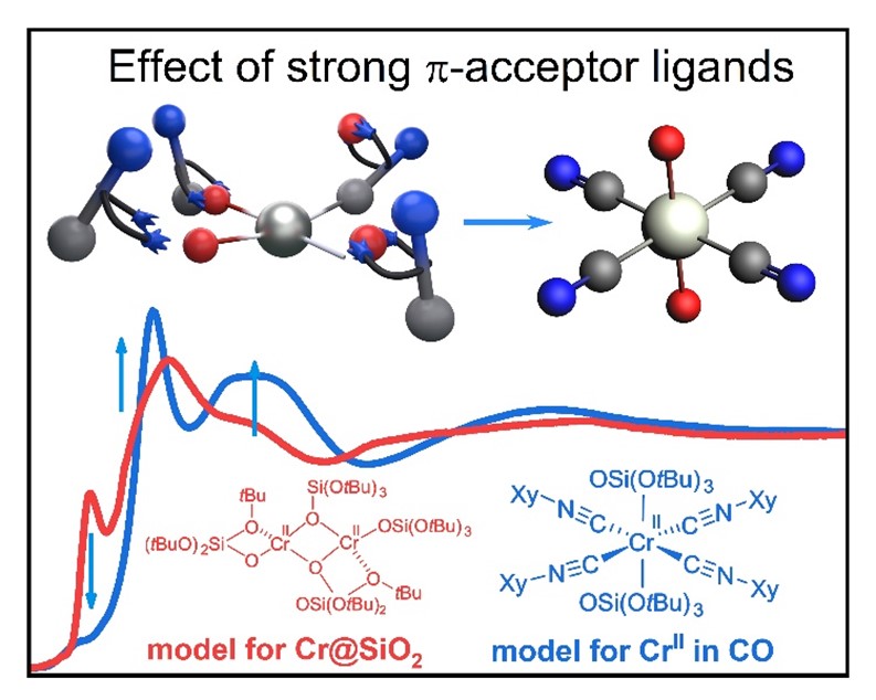
Using the same approach, we (A. Ashuiev et al.) have also characterized the active sites in Cr(III)-based ethylene polymerization catalysts and were able to track Cr(III) alkyl species as propagating species. [5] We (Y. Kakiuchi et al.) have also shown how the presence of strong pi-acceptor ligands like carbon monoxide, a typical probe molecule used to characterize heterogeneous catalysts, can affect the XAS signature and thereby potentially mislead interpretation. All these studies were also made possible by developing a molecular library, here based on isocyanide surrogate molecules, thereby enabling detailed analysis of ligand effects and specific geometries on XAS signatures. [6]

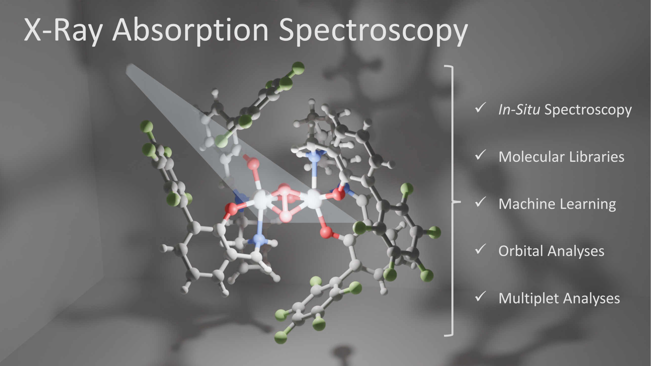
Finally, we (L. Lätsch et al.) have also shown how the less commonly used soft X-rays can provide complementary information to hard X-rays in Ti-based epoxidation catalysts, by exploring the Ti L2,3-edge NEXAFS signatures of key reaction intermediates, e.g. mono- and dinuclear Ti-peroxos or related titanyl functionalities.[7]
Selected References
[1] D. Lebedev et al., J. Am. Chem. Soc. 2018, 140, 451–458
[2] D. Lebedev et al., ACS Cent. Sci. 2020, 6, 7, 1189–1198
[3] S. Docherty et al., J. Am. Chem. Soc. 2023, 145, 25, 13526–13530
[4] D. Trummer et al., J. Am. Chem. Soc. 2021, 143, 19, 7326–7341
[5] A. Ashuiev et al., Angew. Chem. Int. Ed. 2024, 136, e202313348
[6] Y. Kakiuchi et al., Catal. Sci. Technol. 2024, DOI: 10.1039/d3cy01692g
[7] L. Lätsch et al., J. Am. Chem. Soc. 2024, 146, 11, 7456–7466